Combining high-resolution with larger volume images for improved characterization of mudstone reservoirs
Optimization of production from shale reservoirs requires understanding of rock properties over a range of scales.
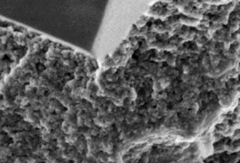
Multiple imaging techniques can be combined to determine the nature, connectivity, and wettability of nano-scale pore systems as well as the underlying mineralogy and organic textures that control reservoir behavior and the propensity of the matrix to fail and to contain expulsion cracks. The current study demonstrates new capabilities in integrated multiscale and time-resolved imaging and analysis workflows for three organic-rich shale samples from two formations. The spatial distributions of connected porosity, organic matter, and microfractures within vertical sub-plugs were quantified from micro-CT imaging, using X-ray contrast enhancement strategies to detect their volume contributions from sub-resolution features, together with tomogram alignment and segmentation. These registered 3D volume distributions comprising billions of voxels showed that most of the porosity in these three samples was hosted by organic matter and most of the coring-induced fractures ran through laminations of locally higher organic content. Dynamic micro-CT imaging was also performed to directly visualize the progress of liquid-liquid diffusion through the pore space. The imaged concentration profiles were fitted to models to estimate the average in-plane diffusivity coefficient. This tomographic analysis was validated and complemented by automated high-resolution 2D back-scattered SEM (BSEM) and SEM-EDS imaging and mapping of pores, organic matter and mineralogy over ion-milled sub-plug sections, and registration of these image mosaics into the corresponding tomogram cross-section. In this way, information on the fine scale of individual features could be combined with statistics over the more representative tomogram volumes. The distribution of organic matter was characterized from this 2D BSEM together with 3D FIBSEM imaging. The majority of organic-hosted connected pores detected by contrast-enhanced micro-CT lay below BSEM and FIBSEM resolution. Secondary electron SEM images (using FESEM) of raw broken surfaces revealed the relatively homogeneous texture of the sub-10 nm pore network permeating the fused aggregates of bitumen nano-granules. Further, the same contrast technique used to highlight bitumen in the tomograms was also applied to ion-milled sections to extend the automated BSEM imaging coupled with SEM-EDS mapping to distinguish bitumen from kerogen at high resolution.