Validation of model predictions of pore-scale fluid distributions during two-phase flow
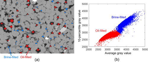
Pore-scale two-phase flow modeling is an important technology to study a rock’s relative permeability behavior. To investigate if these models are predictive, the calculated pore-scale fluid distributions which determine the relative permeability need to be validated. In this work, we introduce a methodology to quantitatively compare models to experimental fluid distributions in flow experiments visualized with microcomputed tomography.

First, we analyzed five repeated drainage-imbibition experiments on a single sample. In these experiments, the exact fluid distributions were not fully repeatable on a pore-by-pore basis, while the global properties of the fluid distribution were. Then two fractional flow experiments were used to validate a quasistatic pore network model. The model correctly predicted the fluid present in more than 75% of pores and throats in drainage and imbibition. To quantify what this means for the relevant global properties of the fluid distribution, we compare the main flow paths and the connectivity across the different pore sizes in the modeled and experimental fluid distributions. These essential topology characteristics matched well for drainage simulations, but not for imbibition. This suggests that the pore-filling rules in the network model we used need to be improved to make reliable predictions of imbibition. The presented analysis illustrates the potential of our methodology to systematically and robustly test two-phase flow models to aid in model development and calibration.