Pore-backs segmentation in PerGeos
Imagery produced from FIBSEMs (Dualbeams) provides users with unique data in terms of resolution and sample size that are not available with other techniques. Despite the power of this analytical approach, some limitations in quantifying the textural data restrict the accuracy of data obtained from FIBSEM images.
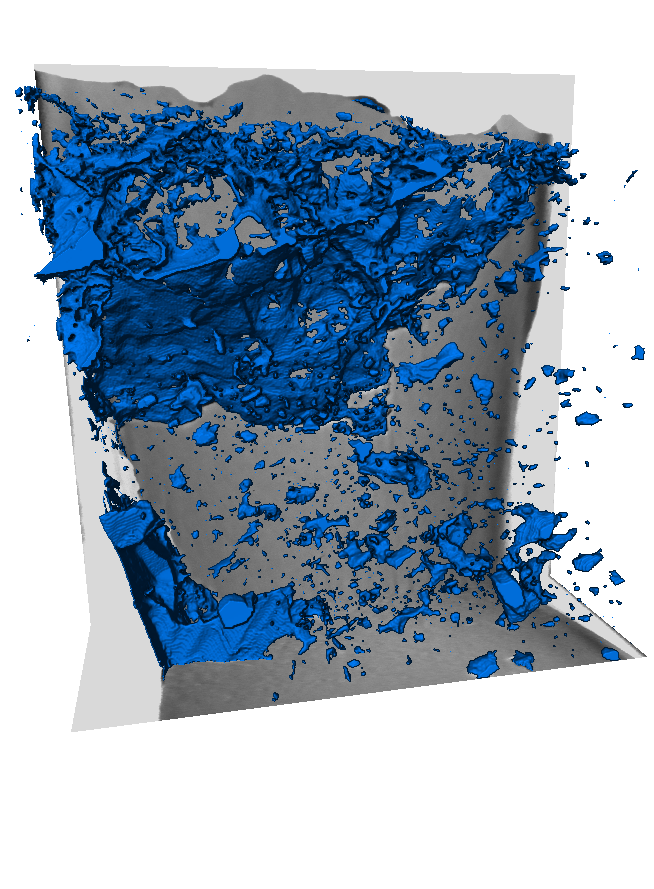
These limitations are derived from artifacts that occur during the acquisition of the data. Collecting data using a FIBSEM is a destructive, serial sectioning technique. A thin slice of material is milled away, the resulting surface imaged, and the process repeated until the requested volume is generated. However, porous materials, by their very nature, create some issues in how to interpret the data that are produced. When each slice is imaged, the empty space (pores and large cracks) are included in the images. If the pore is not deep enough to trap electrons forming a “black” pore synonymous with empty space in SEM imaging, or if it has been filled with redeposited material as a result of the milling process, the back of the pore or other pore filling materials will be imaged. These “pore backs” often termed “pore shine” or “shine through” artifacts make defining the true area of a pore in 2D (volume in 3D) extremely difficult to quantify during post processing using image analysis software.