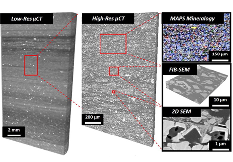
Multi-scale multi-dimensional microstructure imaging of oil shale pyrolysis using X-ray micro-tomography and Electron Microscopy

The complexity of unconventional rock systems is expressed both in the compositional variance of the microstructure and the extensive heterogeneity of the pore space. Visualizing and quantifying the microstructure of oil shale before and after pyrolysis permits a more accurate determination of petrophysical properties which are important in modeling hydrocarbon production potential. We characterize the microstructural heterogeneity of oil shale using X-ray micro-tomography (µCT), automated ultra-high resolution scanning electron microscopy (SEM), MAPS Mineralogy (Modular Automated Processing System) and Focused Ion Beam Scanning Electron Microscopy (FIB-SEM).